


Endocannabinoid System ECS - Introduction and Mechanism 2026
CB1 and CB2 receptors, anandamide and 2-AG, FAAH and MAGL enzymes, and retrograde signaling. What original studies confirm and what remains a hypothesis.
Anandamide, the first endogenous ligand of cannabinoid receptors, was isolated only in 1992 by a team including Devane and Hanus (Devane, Science, 1992). The name of the entire system thus derives from the plant, not the function: researchers first described the receptor to which THC binds, and only later the molecule produced by the body. This guide shows what original studies say about this system: where receptors are located, how the two main endocannabinoids differ, how enzymes terminate the signal, and how retrograde transmission in synapses works. We separately highlight claims about cannabidiol and the entourage effect that lack the support usually attributed to them in cited sources.
KEY INFORMATION
• CB2 gene expression in immune tissues is 10-100 times higher than CB1 (Galiegue, Eur J Biochem, 1995).
• MAGL accounts for about 85% of 2-AG degrading activity in mouse brain (Blankman, Chemistry and Biology, 2007).
• Cannabidiol acts as a negative allosteric modulator at CB1 in cell cultures (Laprairie, BJP, 2015).
• The 2015 Ibeas Bih review considers CBD action through this system unlikely (Neurotherapeutics, 2015).
• Synergy of phytocannabinoids with terpenes remains a hypothesis requiring proof according to Russo (BJP, 2011).
What is the composition of the endocannabinoid system?
This system has three layers: receptors, endogenous lipid ligands, and enzymes that produce and degrade these ligands. Mackie describes it exactly in this composition in a 2008 review, noting that many cannabinoid effects are mediated through two G protein-coupled receptors, CB1 and CB2, although other proteins may also be involved (Mackie, Journal of Neuroendocrinology, 2008). Both receptors primarily couple to inhibitory G proteins and follow pharmacological principles common to this receptor family. Mackie lists three important features for cellular response to a ligand: partial agonism, functional selectivity, and inverse agonism. This explains why two substances binding the same receptor can have opposite effects.
A feature distinguishing this network from classical neurotransmission is the lack of storage. Dopamine and serotonin wait in synaptic vesicles for release signals. Endocannabinoids are produced only when needed, from membrane lipids, and disappear quickly by enzymatic breakdown. Thus, the signal is limited in time and space to the immediate vicinity of its origin. Wilson and Nicoll showed in 2001 that depolarized hippocampal neurons release anandamide and 2-AG in a calcium-dependent manner (Wilson and Nicoll, Nature, 2001).
The table below summarizes the core of this network along with the study that first described each element. This is not a complete list of lipid signaling proteins, only those for which original work is cited later in the text.
| Layer | Element | Describing Study |
|---|---|---|
| receptor | CB1 | Matsuda, Nature, 1990 |
| receptor | CB2 | Munro, Nature, 1993 |
| ligand | anandamide (AEA) | Devane, Science, 1992 |
| ligand | 2-arachidonoylglycerol (2-AG) | Mechoulam, 1995 |
| degrading enzyme | FAAH | Cravatt, Nature, 1996 |
| degrading enzyme | MAGL | Dinh, PNAS, 2002 |
What was the discovery history from 1964 to 2002?
Less than forty years passed from THC isolation to description of the enzyme degrading the second endocannabinoid. Russo himself gives the starting point: tetrahydrocannabinol has been the main subject of cannabis research since 1964, when Raphael Mechoulam isolated and synthesized it (Russo, British Journal of Pharmacology, 2011). The next two decades were spent finding where this molecule acts. Progress came first from synthesizing potent THC derivatives, then from cloning the receptor gene.
In 1988, Devane and colleagues identified and characterized the cannabinoid binding site in rat brain using tritiated CP-55,940. The dissociation constant was 133 pM, and binding was saturable, stereoselective, and sensitive to a non-hydrolyzable GTP analog, indicating G protein regulation (Devane, Molecular Pharmacology, 1988). Two years later, Matsuda et al. reported the receptor structure and cloned cDNA expression (Matsuda, Nature, 1990).
In 1993, the second receptor was discovered. Munro, Thomas, and Abu-Shaar cloned a protein absent in the brain but present in marginal zone spleen macrophages (Munro, Nature, 1993). Ligands appeared in parallel: anandamide in 1992, and in 1995 a monoacylglycerol from dog intestine binding cannabinoid receptors (Mechoulam, Biochemical Pharmacology, 1995). Enzymes completed the picture: fatty acid amide hydrolase in 1996 (Cravatt, Nature, 1996) and brain monoacylglycerol lipase in 2002 (Dinh, PNAS, 2002).
Where in the brain are CB1 receptors located?
CB1 receptors are found in very high amounts in several brain regions and at lower concentrations diffusely throughout the central nervous system. They mediate the psychoactive effects of cannabinoids, while CB2 has a much narrower distribution, mainly on immune cells and a few neurons (Mackie, Journal of Neuroendocrinology, 2008). The popular comparison that CB1 density exceeds mu opioid receptor density by a hundredfold does not come from this work and was removed from the text.
Receptor localization in synapses reveals more than numbers. Wilson and Nicoll showed that in the hippocampus, CB1 is mainly expressed in inhibitory GABAergic interneurons and concentrated on axon terminals, i.e., presynaptic side. A synthetic agonist reduced gamma-aminobutyric acid release in hippocampal slices (Wilson and Nicoll, Nature, 2001). Thus, the receptor acts as a brake on neuron output, not as a transmitter of its own signal. Galiegue et al. add peripheral expression: CB1 transcripts were detected in adrenal gland, heart, lung, prostate, uterus, ovary, testis, bone marrow, thymus, and tonsils, though at much lower levels than in the brain.
This localization explains why the same receptor is sometimes described as silencing excitation and sometimes as enhancing it. If CB1 is on inhibitory neuron terminals, its activation removes inhibition from the target cell. If on excitatory terminals, the effect is opposite. The direction depends on the cell population studied. More on the distribution of both receptors is in a separate guide on CB1 and CB2 receptors.
What role do CB2 receptors play in immunity?
The strongest measurement of CB2 distribution comes from Galiegue et al. 1995. The team quantitatively measured transcripts of both receptors in human immune tissues and cells by PCR. The CB2 gene is not expressed in the brain, but in immune tissues its level was 10-100 times higher than CB1 (Galiegue, European Journal of Biochemistry, 1995). In spleen and tonsils, CB2 mRNA levels matched CB1 mRNA levels in the central nervous system. The CB2 expression dominance in immune tissues was independently confirmed by Northern blot.
The study also provides a cell order often misquoted. CB2 mRNA levels rank as follows: B lymphocytes highest, then NK cells, followed by monocytes, neutrophils, CD8 T cells, and finally CD4 T cells. The 10-100 fold figure describes CB2 dominance over CB1 in immune tissues, not B cells over T cells. Antibody staining confirmed CB2 protein presence in B cell-rich areas. The same pattern appeared in human myeloid, monocytic, and lymphoid cell lines.
| Common claim in popular texts | What Galiegue 1995 states |
|---|---|
| CB2 is 10-100 times more abundant in B lymphocytes than T lymphocytes | CB2 is 10-100 times more abundant than CB1 in immune tissues |
| CB2 is absent outside the immune system | CB2 mRNA was also detected in other peripheral tissues but at very low levels |
| CB1 is exclusively a brain receptor | CB1 was detected in several peripheral tissues, though at much lower levels than in the brain |
What happens after CB2 receptor activation?
Receptor distribution alone does not indicate the effect of its activation. A 2009 review by Nagarkatti et al. on cannabinoids as a new class of anti-inflammatory drugs provides answers. The starting point is the same as Galiegue’s measurement: CB1 is mainly expressed in the brain, CB2 mainly on immune cells, and presence of both receptors on these cells suggests cannabinoids regulate immunity (Nagarkatti, Future Medicinal Chemistry, 2009).
The authors summarize three mechanisms from experimental studies. THC administration to mice induced pronounced apoptosis of T lymphocytes and dendritic cells, weakening immunity. In several models, cannabinoids reduced cytokine and chemokine production and increased regulatory T cells, suppressing inflammation. The third line concerns endocannabinoids themselves: their administration or inhibition of degrading enzymes weakened immunity and reversed immune-mediated organ damage, including in the liver.
The anti-inflammatory profile of cannabinoids was described even before the second receptor. Munro et al. 1993 list THC effects unrelated to psychoactivity: analgesic, anti-inflammatory, immunosuppressive, anticonvulsant, lowering intraocular pressure in glaucoma, and antiemetic (Munro, Nature, 1993). Finding a receptor absent in the brain gave these observations a site of action. Note that most data come from animal models and cell cultures, not human studies.
Is there a third cannabinoid receptor?
The question of CB3 receptor recurs in texts about GPR55 and TRPV1, and the pharmacological community’s answer since 2010 is consistent. The International Union of Basic and Clinical Pharmacology review lists criteria any new cannabinoid receptor must meet and states that currently no receptor or channel outside CB1 and CB2 meets them (Pertwee, Pharmacological Reviews, 2010).
This does not mean there are no candidates. The same review points to targets worth further study, including the vanilloid receptor TRPV1, which may act as an ionotropic cannabinoid receptor under physiological or pathological conditions, and some orphan G protein-coupled receptors. The authors also discuss CB1’s ability to form heteromeric complexes with other family receptors and existence of several uncharacterized cannabinoid receptors. The review covers orthosteric and allosteric interactions of cannabinoid ligands with recognized receptors beyond CB1 and CB2, orphan receptors, ligand-gated ion channels, transient receptor potential channels, and nuclear receptors activated by peroxisome proliferators. A separate chapter addresses current nomenclature.
The practical takeaway: the name CB3 in marketing materials is a shorthand, not an established pharmacological entity. Ligands acting similarly at CB1 or CB2 may have distinct pharmacological profiles because they also target other proteins.
How does anandamide differ from 2-AG?
The two main endocannabinoids differ not only in structure but also in receptor activation strength. Sugiura’s 2006 review states anandamide usually acts as a partial agonist at cannabinoid receptors, while 2-AG fully activates them in most systems (Sugiura, Progress in Lipid Research, 2006). Structure-activity studies led authors to conclude 2-AG, not anandamide, is the natural ligand for both receptors. The difference also concerns quantity: 2-AG is present in the brain at concentrations 170 times higher than anandamide, measured by Stella et al. (Nature, 1997). The rounded figure of 200 circulating in literature is attributed to Sugiura’s review, which does not state it.
2-AG formation is rapid and depends on phospholipid metabolism containing arachidonic acid, especially increased turnover of inositol phospholipids upon cell stimulation. The same study assigns 2-AG a role as a retrograde messenger regulating synaptic transmission and involvement in inflammation and immune response. Anandamide was isolated earlier, in 1992, as a brain component binding cannabinoid receptors (Devane, Science, 1992). Sugiura notes 2-AG was independently described in 1995 in rat brain and dog intestine. The latter location is often omitted but explains why this system is now discussed beyond neurology.
Popular numbers describing concentrations, such as 200-fold 2-AG excess over anandamide or 30-50 pmol per gram tissue range, do not appear in the abstract and were removed. We retained what the source states directly: the difference in agonism type. Separate discussions of both molecules are in guides on 2-AG and anandamide.
How do FAAH and MAGL enzymes terminate the signal?
Endocannabinoid signaling ends by enzymatic degradation, and enzymes, not receptors, determine signal duration. Cravatt et al. characterized the enzyme breaking down neuromodulatory fatty acid amides, FAAH hydrolase, in 1996 (Cravatt, Nature, 1996). Six years later, Dinh et al. cloned rat brain monoacylglycerol lipase and showed its hydrolysis is the main mechanism inactivating 2-AG in intact neurons (Dinh, PNAS, 2002).
Blankman measured the division of labor. Functional proteomics mapped 2-AG degrading enzymes in mouse brain: about 85% of activity is MAGL, the remaining 15% mainly two previously undescribed proteins, ABHD6 and ABHD12 (Blankman, Chemistry and Biology, 2007). All three enzymes have different intracellular distributions, suggesting control of distinct pools of the same molecule.
MAGL mRNA distribution in rat brain is uneven, highest where CB1 receptor is present: hippocampus, cortex, anterior thalamus, and cerebellum. Immunohistochemistry in hippocampus showed clear laminar distribution, indicating presynaptic localization. The enzyme is exactly where the molecule it degrades ends. Dinh et al. also showed functional effect: introducing extra copies of the enzyme gene into rat cortex neurons via adenovirus increased expression and reduced 2-AG accumulation after stimulation. The same change did not affect anandamide accumulation, confirming the division of labor between enzymes.
What is retrograde signaling in synapses?
Classically, synaptic information flows one way: presynaptic neuron releases neurotransmitter, postsynaptic neuron receives it. Wilson and Nicoll showed in 2001 that transient weakening of GABAergic transmission after depolarization of hippocampal pyramidal cells is mediated by a signal running backward via release of endogenous cannabinoids (Wilson and Nicoll, Nature, 2001). The neuron thus gains a mechanism to communicate retrogradely and modulate its own inputs.
The sequence is: depolarization opens voltage-dependent calcium channels, intracellular calcium rise triggers endocannabinoid synthesis from membrane lipids, the molecule crosses the synaptic cleft to presynaptic terminal, binds CB1, and reduces neurotransmitter release. Sugiura identifies 2-AG as the messenger fulfilling this role (Sugiura, Progress in Lipid Research, 2006).
This mechanism’s significance goes beyond physiological curiosity. Since the receiving cell can reduce signal strength at the source, it has a local feedback loop operating at single synapses without higher centers. This explains why CB1 agonists affect short-term memory and time perception, functions dependent on precise hippocampal network rhythms. Note the discovery order: first it was known that depolarized hippocampal neurons release both endocannabinoids calcium-dependently and that synthetic CB1 agonists reduce GABA release. The 2001 study connected these facts into one mechanism.
What is the clinical endocannabinoid deficiency theory?
This hypothesis was formulated by Ethan Russo, with a review published in 2016 (Russo, Cannabis and Cannabinoid Research, 2016). The premise is that every person has a baseline endocannabinoid activity level, determined by anandamide and 2-AG concentrations, their production and degradation rates, and receptor number and state. The theory states that in certain congenital or acquired conditions, this level drops and itself causes symptoms.
Russo identifies three syndromes: migraine, fibromyalgia, and irritable bowel syndrome. They share lack of clear routine diagnostic changes, resistance to multiple drugs, overlapping patient populations, and a common pattern of pain hypersensitivity with central sensitization features. The author recalls that patients with these diagnoses were long labeled psychosomatic.
Important is what changed between the first hypothesis formulation and the 2016 review. Russo writes that initially, the hypothesis was based on genetic overlap, comorbidity, and observation that cannabinoid treatment often brought relief, but lacked objective proof and formal clinical data. Today, statistically significant differences in cerebrospinal fluid anandamide levels in migraine patients are documented, and imaging shows reduced system activity in PTSD. This supports the hypothesis but does not confirm it.
Does CBD really act through the endocannabinoid system?
This question has two correct answers that must be presented side by side, as either alone is misleading. Laprairie et al. showed in 2015 that cannabidiol reduces efficacy and potency of 2-AG and THC on CB1-dependent pathways in HEK 293A cells and striatal neuron models. It also reduced arrestin 2 binding to the receptor, preventing internalization. They call this non-competitive negative allosteric modulation (Laprairie, British Journal of Pharmacology, 2015).
The second answer comes from the 2015 review by Ibeas Bih et al. They surveyed molecular pharmacology literature on cannabidiol and counted over 65 distinct molecular targets. Their conclusion differs from the number: current evidence indicates cannabidiol does not directly interact with the endocannabinoid system outside of cell culture and supraphysiological concentrations (Ibeas Bih, Neurotherapeutics, 2015). In neurological diseases, action through this system is considered very unlikely.
There is no contradiction. Laprairie describes molecule behavior in culture; Ibeas Bih assesses whether this behavior translates to the organism at achievable oral doses, which are low due to poor absorption. Claims like “CBD raises endogenous endocannabinoid levels by inhibiting FAAH” lack support in these sources and were removed. After excluding unlikely targets, the review retains those related to intracellular calcium regulation, including VDAC1 channel, GPR55 receptor, and T-type calcium channels.
What does the entourage effect study really say?
The entourage effect is presented in marketing as justification for choosing full-spectrum extract over isolate. The most cited study says something more cautious. Russo discusses cannabis terpenoids, their shared precursor with phytocannabinoids, and their status as generally recognized as safe substances, then points to interactions that could produce synergy in pain, inflammation, depression, anxiety, addiction, epilepsy, and cancer (Russo, British Journal of Pharmacology, 2011).
The concluding sentence is conditional: synergy of phytocannabinoids with terpenoids, if proven, increases the likelihood that this plant can yield a broad line of new therapeutic products. The author proposes methods to study the entourage effect in future experiments, indicating the issue was open then. The study is a single-author review, not a comparison of extract versus isolate.
One interaction was directly measured. Gertsch et al. showed beta-caryophyllene, a volatile component of essential oils from many culinary and spice plants, selectively binds CB2 receptor with affinity constant 155 nM and acts as a functional agonist (Gertsch, PNAS, 2008). In CB2 knockout mice, the anti-inflammatory effect of this terpene disappeared, indicating the receptor as the site of action. In cannabis, it is a major component. Upon CB2 binding, it inhibited adenylate cyclase, caused transient intracellular calcium changes, and reduced lipopolysaccharide-induced proinflammatory cytokine production in peripheral blood. This is the only terpene family member with a directly measured site of action.
How does Polish law define the THC threshold?
Phytocannabinoids in Poland are regulated by a law directly referring to the content of two substances, not one. Industrial hemp are Cannabis sativa L. plants in which the sum of delta-9-THC and tetrahydrocannabinolic acid (THCA) in flowering or fruiting tops, from which resin has not been removed, does not exceed 0.3% dry weight. The sum is rounded to one decimal place.
The basis is Article 4 point 5 of the Act of July 29, 2005 on counteracting drug addiction (consolidated text Journal of Laws 2023 item 1939), as amended by the Act of March 24, 2022 (Journal of Laws 2022 item 763), effective May 7, 2022. The August 27, 2026 amendment did not change this provision.
Two things should be distinguished. First, the threshold counts the sum of delta-9-THC and THCA, not delta-9-THC alone, which changes laboratory test results for the same sample. Second, the national threshold corresponds to the EU threshold in value but does not derive from it. Varieties qualifying for support under the Common Agricultural Policy may contain up to 0.3% THC from January 1, 2023 based on Article 4 paragraph 4 of Regulation (EU) 2021/2115; previously the EU threshold was 0.2%. These are two separate regulations with the same number. Neither the amending act nor its counterpart cites the EU regulation as their basis. The correct statement is: the national threshold corresponds to the EU threshold. Saying the Polish threshold derives from the EU regulation is incorrect despite the numbers matching.
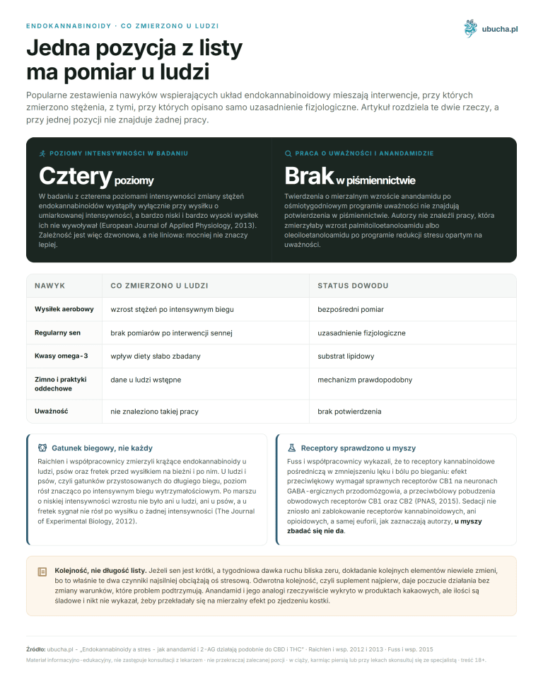
Does physical activity change endocannabinoid levels?
Yes, and this is one of the few observations in this area made in humans. Sparling et al. studied trained students running on a treadmill or cycling for 50 minutes at 70-80% maximum heart rate. The study provides the first evidence that moderate intensity exercise stimulates the endocannabinoid system and suggests it as a possible mechanism for exercise-induced analgesia and other training adaptations (Sparling, Neuroreport, 2003).
Three numbers often cited with this study do not appear in the abstract: twofold increase in blood anandamide, 60-70% maximum heart rate as optimal, and attenuation above 80%. The actual range used was 70-80%, and only this value should be cited. Also note the group: trained students, not inactive individuals, so extrapolating to beginners is an assumption not confirmed by this study.
A second line concerns genetics. Dincheva et al. created a mouse with a human FAAH gene variant C385A (rs324420). This variant alters enzyme expression and activity, raising anandamide levels. Carriers, both mice and humans, had reduced FAAH expression, stronger functional connectivity between prefrontal cortex and amygdala, more efficient fear extinction, and fewer anxiety behaviors (Dincheva, Nature Communications, 2015). The study appeared in Nature Communications, not PNAS as previously stated.
Which organisms have an endocannabinoid system?
The answer is less uniform than popular summaries suggest. McPartland et al. searched genomes of twelve evolutionarily distant organisms, from humans and mice, through Japanese pufferfish, sea anemone, nematode, and fruit fly, to yeast, Arabidopsis, malaria parasite, ciliate, and two microorganisms. Orthologs were identified by phylogenetic methods, and their functional potential predicted based on amino acid motifs known to participate in these proteins’ actions (McPartland, Gene, 2006).
The result showed individual genes have different evolutionary paths. Orthologs of TRPV1 and GPR55 are limited to mammals. CB2 receptor and DAGL beta appear in vertebrates. MAGL and COX2 reach chordates. DAGL alpha and CB1-like receptors occur in animals. NAPE-PLD is found in animals and fungi. FAAH extends to all eukaryotes. There is no single date from which the entire system exists.
The authors note their method detected fewer orthologs than automatic annotation systems, so the result is cautious rather than inflated. The novelty was overlaying protein function information on gene trees, allowing assessment not only of sequence similarity but also the likelihood that an ortholog performs the same role. The discussion covers phylogenetic profiles, non-orthologous gene displacement, convergent function, and coevolution. The statement that CB1 and CB2 genes have been conserved for over 600 million years does not appear and was removed. A broader comparison of compounds produced by organism and plant is in the text on phytocannabinoids and endocannabinoids.
Frequently Asked Questions
What is the endocannabinoid system?
It is a lipid signaling network composed of endocannabinoids, cannabinoid receptors, and enzymes that produce and break down these ligands. Many cannabinoid effects are mediated through two G protein-coupled receptors, CB1 and CB2, although other proteins may also be involved (Mackie, Journal of Neuroendocrinology, 2008).
Where are CB1 and CB2 receptors located?
CB1 is found in very high amounts in several brain regions and at lower concentrations dispersed elsewhere. CB2 has a narrower distribution and is mainly present on immune cells. The CB2 gene is not expressed in the brain, and in immune tissues its level is 10-100 times higher than CB1 (Galiegue, European Journal of Biochemistry, 1995).
How does anandamide differ from 2-AG?
Anandamide usually acts as a partial agonist at cannabinoid receptors, whereas 2-AG fully activates them in most systems. Structure-activity relationship studies led the authors to conclude that 2-AG is more likely the natural ligand for both receptors (Sugiura, Progress in Lipid Research, 2006).
How do FAAH and MAGL enzymes work?
FAAH breaks down neuromodulatory fatty acid amides, including anandamide (Cravatt, Nature, 1996). MAGL inactivates 2-AG and accounts for about 85% of the 2-AG degrading activity in the mouse brain, with the remaining 15% mainly attributed to ABHD6 and ABHD12 proteins (Blankman, Chemistry and Biology, 2007).
What is the clinical endocannabinoid deficiency theory?
This is Ethan Russo’s hypothesis that a lowered baseline activity of this system itself causes symptoms of migraine, fibromyalgia, and irritable bowel syndrome. It is supported by documented differences in anandamide levels in cerebrospinal fluid of migraine sufferers (Russo, Cannabis and Cannabinoid Research, 2016).
Does CBD act through the endocannabinoid system?
In cell cultures, cannabidiol behaves as a non-competitive negative allosteric modulator of CB1 (Laprairie, British Journal of Pharmacology, 2015). However, the 2015 review by Ibeas Bih considers direct interaction with this system outside of supraphysiological concentrations in culture unlikely (Neurotherapeutics, 2015).
What is retrograde signaling?
The signal runs opposite to classical synapses, from the receiving cell back to the terminal releasing the neurotransmitter. Transient weakening of GABAergic transmission after depolarization of hippocampal pyramidal cells is mediated by released endocannabinoids (Wilson and Nicoll, Nature, 2001). This allows the neuron to modulate its own synaptic inputs.
Do animals have an endocannabinoid system?
Individual genes have different evolutionary paths. CB1-like receptors and DAGL alpha occur in animals, CB2 and DAGL beta in vertebrates, MAGL in chordates, and FAAH even in eukaryotes. Orthologs of TRPV1 and GPR55 are limited to mammals (McPartland, Gene, 2006).
Which claims about this system lack support in sources?
While organizing this article, six identifiers led to studies in completely different fields, including a study on chromosome breakpoints in T cell tumors and research on insulin-like growth factor receptor. Several numbers did not appear in the cited works. The table below compares frequently repeated statements with what the cited source actually says.
| Repeated claim | What the cited source states |
|---|---|
| 2-AG concentration in brain is about 200 times higher than anandamide | The number is close but attributed to Stella et al. (Nature, 1997) as 170 times, not 200. Sugiura’s 2006 review describes the difference in agonism type. |
| CB1 density exceeds mu opioid receptor density by 100 times | Mackie 2008 reports very high CB1 amounts in several brain regions without opioid system comparison. |
| CB1 and CB2 genes have been conserved for over 600 million years | McPartland 2006 shows different evolutionary paths for individual genes, no single date. |
| Running doubles anandamide, optimum at 60-70% max heart rate | Sparling 2003 studied exercise at 70-80% max heart rate and does not report fold increase. |
| FAAH C385A variant reduces enzyme activity by 50% in 20% of Europeans | Dincheva 2015 reports reduced enzyme expression and higher anandamide without these percentages. |
| Full-spectrum extract acts stronger than isolate due to proven entourage effect | Russo 2011 states synergy conditionally and proposes methods to study it. |
| CBD raises endocannabinoid levels by inhibiting FAAH | Ibeas Bih 2015 considers direct CBD action through this system unlikely. |
The common denominator is that identifiers led to existing and usually relevant studies, so address-level checks passed. Discrepancies appear only when comparing statements with abstracts, requiring opening the study. Therefore, each number in this article is accompanied by the first author’s name, journal, and year, with links to the study record, not the publisher’s homepage. Readers can verify each in seconds.
Summary
The collected studies form a coherent picture, narrower than that circulating in popular texts. Certain are the existence of two receptors with distinct distribution, two endogenous ligands differing in agonism type, two degrading enzymes, and retrograde synaptic signaling. Each element has an original describing study, verifiable by the given identifier.
Much less certain is external control of this system. The endocannabinoid deficiency theory remains a hypothesis with support but no resolution. Phytocannabinoid-terpenoid synergy is conditionally proposed by Russo. Direct cannabidiol action through this system is considered unlikely at achievable concentrations by the 2015 review. These three statements should be kept together, as marketing materials often present them without caveats. A warning sign appears already in the 1988 study: in binding tests, cannabidiol and cannabigerol displaced labeled ligand from the receptor at less than half the efficacy of THC and its hydroxyl derivative at 1 micromolar (Devane, Molecular Pharmacology, 1988). Cannabidiol’s weak affinity for this receptor is not a recent discovery.
We removed six incorrect identifiers leading to unrelated studies and every number absent from cited sources. If you seek products containing phytocannabinoids, their selection is in the oils category.
This article is for informational and educational purposes and does not constitute medical advice. Consult a physician before starting cannabis or CBD for therapeutic purposes, especially if taking other medications, pregnant, or breastfeeding.
Author: Michał Waluk · Published: 2026-05-11 · Updated: 2026-08-10