


2-AG: the most abundant endocannabinoid (guide)
2-AG is present in the brain at a concentration 170 times higher than anandamide and is a full CB1 agonist. What has been measured in animals, and what has not been checked in humans.
When it comes to endocannabinoids, anandamide gets most of the attention. Meanwhile, 2-arachidonoylglycerol, or 2-AG, is found in the brain at a concentration 170 times higher and fully activates cannabinoid receptors (Stella et al., Nature, 1997). It was discovered in 1995 by two independent teams working on two different tissues in two different countries. This article explains what 2-AG is, how it is produced, and what it does, but it also keeps an eye on something that guides to the endocannabinoid system usually overlook: it provides research material with every finding. Because almost everything we know about this molecule has been measured on slices of rodent brains or experimental animals, not on humans.
KEY INFORMATION
• In the brain, 2-AG is present at levels 170 times higher than anandamide and acts on cannabinoid receptors as a full agonist (Stella et al., Nature, 1997).
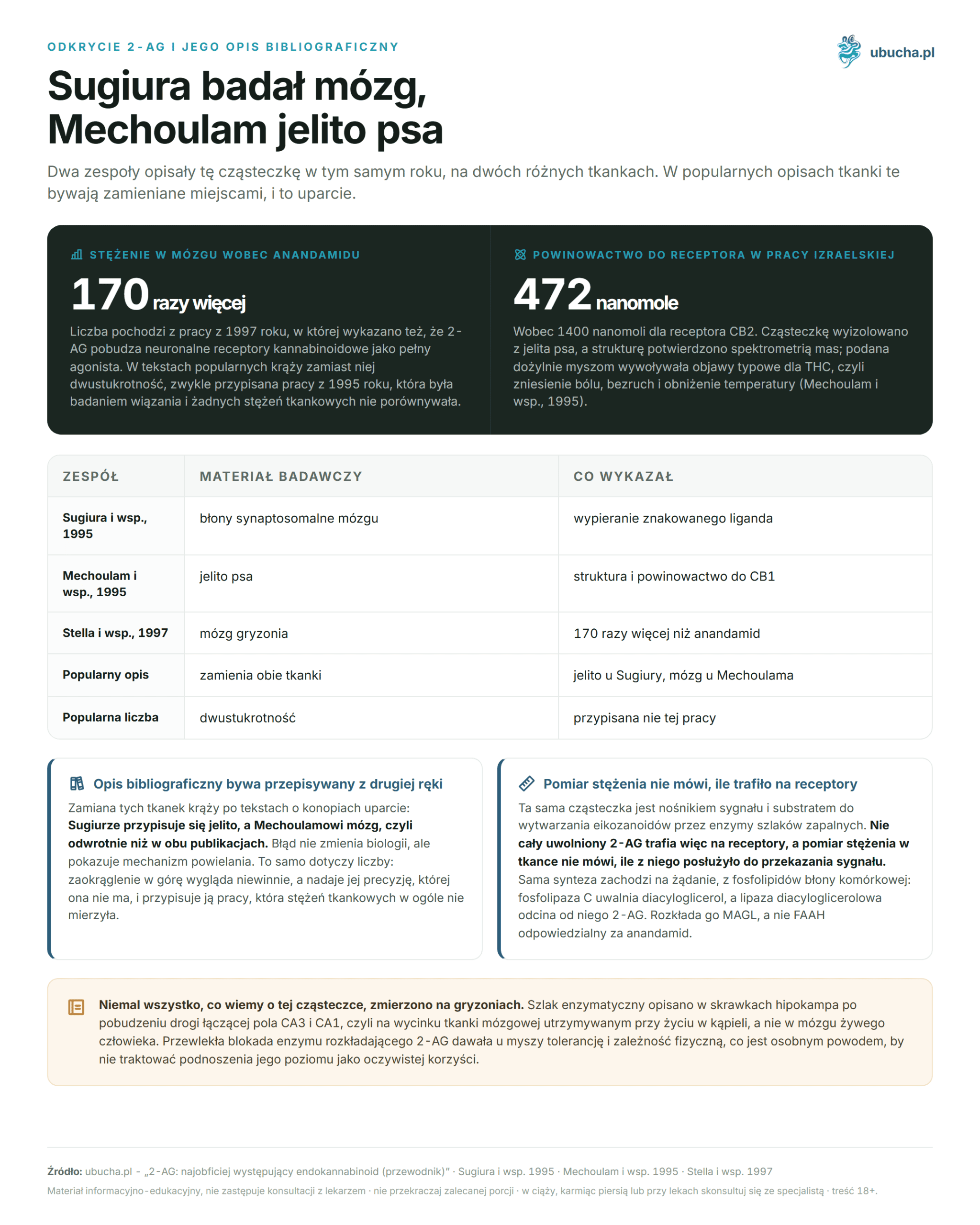
• The Sugiura team isolated 2-AG from the brain, while the Mechoulam team isolated it from dog intestine.
• It is broken down by MAGL, not FAAH responsible for anandamide.
• Chronic MAGL blockade in mice led to tolerance and physical dependence.
What is 2-AG and where does it come from?
2-arachidonoylglycerol is an ester of arachidonic acid and glycerol. Like all endocannabinoids, it is not stored in vesicles but is produced on demand when a neuron is stimulated. Synthesis occurs from membrane phospholipids in two steps: phospholipase C releases diacylglycerol, and diacylglycerol lipase cleaves 2-AG from it. Two variants of this second enzyme, designated alpha and beta, have different distributions in tissues.
The formation of 2-AG depends on calcium, and its enzymatic pathway has been described in slices of the hippocampus after stimulation of the pathway connecting CA3 and CA1 fields. This is an important methodological detail: the mechanism was established on a slice of rodent brain kept alive in a bath, not in the brain of a living human.
2-AG concentrations in the brain are high, but their reading can be misleading. The same molecule serves as a signal carrier and a substrate for the production of eicosanoids by enzymes of inflammatory pathways. Therefore, not all released 2-AG reaches receptors, and measuring the concentration in tissue does not indicate how much of it was used for signaling. We discuss the entire system more broadly in the introduction to the endocannabinoid system.
Who discovered 2-AG and from what material?
Two teams, in the same year, on two different tissues, and it is worth distinguishing them, as they are often confused in popular descriptions. The Sugiura team in Japan studied binding to synaptosomal membranes, which is a preparation from nerve endings of the brain. They showed that 2-AG displaces a labeled ligand from the cannabinoid receptor, with affinity increasing significantly after inhibiting esterases or cooling the sample to zero degrees (Sugiura et al., Biochemical and Biophysical Research Communications, 1995).
The Mechoulam team in Israel isolated the same molecule from dog intestine, confirming the structure by mass spectrometry. The compound bound to the CB1 receptor at a constant of 472 nanomoles and to CB2 at 1400 nanomoles, and when administered intravenously to mice, it produced symptoms typical of THC: pain relief, immobility, and lowered temperature (Mechoulam et al., Biochemical Pharmacology, 1995).
We have noticed that the swapping of these tissues circulates in texts about cannabis persistently: Sugiura is attributed to the intestine, and Mechoulam to the brain, which is the opposite of both publications. The error does not change the biology but shows the mechanism of replication: bibliographic descriptions are often copied second-hand.
How much more 2-AG is there in the brain than anandamide?
One hundred seventy times, and this number has a specific source. Stella, Schweitzer, and Piomelli demonstrated in 1997 that 2-AG is present in the brain at levels 170 times higher than anandamide, stimulates neuronal cannabinoid receptors as a full agonist, and prevents the induction of long-term synaptic potentiation in connections between CA3 and CA1 fields of the hippocampus (Stella et al., Nature, 1997).
In popular texts, instead, the number of two hundred times circulates, usually attributed to Sugiura’s work from 1995. This is a double inaccuracy. Sugiura’s work was a binding study and did not compare any tissue concentrations, while the value measured by another team two years later is 170, not 200. Rounding up looks innocent, but it gives the number a precision it does not have.
The research material also needs to be mentioned here, because without it, the number sounds like a biological constant. The measurement concerned rodent brain tissue, not the human brain. There is no reason to believe that the advantage of 2-AG over anandamide in humans disappears, but the number itself describes an animal.
How does 2-AG differ from anandamide?
There are several differences concerning the type of agonism, concentrations, enzymes, and how each reacts to cannabidiol. The table below compares them along with the material on which each was established.
| Feature | 2-AG | Anandamide (AEA) |
|---|---|---|
| Year of description | 1995 | 1992 |
| Concentration in rodent brain | 170 times higher | Reference point |
| Type of CB1 agonist | Full agonist | Partial agonist |
| Synthesis enzyme | Diacylglycerol lipase | NAPE-PLD |
| Degradation enzyme | MAGL | FAAH |
| Effect of chronic enzyme blockade in mice | Tolerance, physical dependence, CB1 desensitization | Lasting pain relief without CB1 desensitization |
| CBD effect | Undocumented | Moderate inhibition of degradation, increased serum levels in humans |
The last two rows are the most important part of this comparison and also the part that usually does not appear in popular tables. Both endocannabinoids behave completely differently under chronic pharmacological intervention.
The difference between a full and a partial agonist is not just a formality. A full agonist produces a maximum receptor response, while a partial agonist stops below that threshold even with full saturation of binding sites. Therefore, 2-AG is considered the main signal carrier in the synapse, while anandamide is seen as a background regulator. This division has been derived from rodent tissue.
On what was the retrograde signaling of 2-AG measured?
On slices of rodent hippocampus. Wilson and Nicoll demonstrated that the transient weakening of inhibitory transmission that occurs after depolarization of a pyramidal neuron is retrogradely transmitted by endocannabinoids. The stimulated neuron releases them in a calcium-dependent manner, and they bind to the CB1 receptor on the presynaptic terminal and suppress the release of gamma-aminobutyric acid (Wilson and Nicoll, Nature, 2001).
This reverses the usual direction: the cell receiving the signal responds to the sender and asks it to be quieter. The mechanism acts like a safety valve for the neuronal network, which is why it is studied in the context of epilepsy, where the problem is excessive excitation of the network.
One caveat is worth remembering. The 2001 paper speaks of endocannabinoids in the plural because depolarized hippocampal neurons release both anandamide and 2-AG. Attributing the entire phenomenon solely to 2-AG is a conclusion from later literature, not the content of this experiment. In abstracts, this distinction disappears, along with the information that the role of 2-AG itself was established here retroactively, based on works published many years later.
Is blocking the MAGL enzyme a safe way to relieve pain?
Not as safe as the widespread belief suggests about its action being comparable to opioids and devoid of addiction risk. This statement contradicts the work that directly studied chronic MAGL inhibition. After multiple administrations, the inhibitor JZL184 lost its analgesic effect and caused cross-tolerance to CB1 receptor agonists in mice. The same picture emerged from the knockout of the MAGL coding gene (Schlosburg et al., Nature Neuroscience, 2010).
Chronic MAGL blockade also induced physical dependence, impaired endocannabinoid-dependent synaptic plasticity, and desensitized brain CB1 receptors. The authors compared this to FAAH blockade, the enzyme that breaks down anandamide, which provided lasting pain relief without damaging receptors. The conclusion of the work is the opposite of popular belief: the two endocannabinoids provide different analgesic profiles, and raising 2-AG leads to functional antagonism of the system, not its enhancement.
MAGL inhibitors remain a laboratory tool for separating the roles of both endocannabinoids (Blankman and Cravatt, Pharmacological Reviews, 2013). This review describes only preclinical models. We do not provide the number of clinical studies or their dates because there is no basis for any specific value in the reviewed sources.
What has been shown about 2-AG after brain injury?
This has been demonstrated in mice, and the result is clear. After closed head injury, the concentration of 2-AG in brain tissue significantly increased. Administration of synthetic 2-AG after injury reduced brain swelling, improved clinical assessment scores, reduced infarct volume, and limited hippocampal cell death compared to the control group (Panikashvili et al., Nature, 2001).
Two things in this work are often misrepresented. First, the multiplicity of 2-AG increase is sometimes stated as tenfold within minutes, while the abstract only mentions a significant increase. Second, the beneficial effect was dose-dependently weakened by a CB1 receptor antagonist. Thus, CB1 was tested, not CB1 together with CB2, and only about the former can be discussed here.
Interestingly, the administration of 2-AG together with inactive 2-acylglycerols, which are naturally present in the brain, enhanced recovery. The translation of these results to humans remains open, and there is no practical application that could be honestly written about today. The gap between a mouse after head injury and a neurosurgery patient is a rule in this field.
Does CBD raise 2-AG levels?
There is no data on this, and the pathway indicated by studies in humans leads to the second endocannabinoid. In a clinical study of individuals with schizophrenia, treatment with cannabidiol raised serum anandamide levels, and this increase was associated with clinical improvement. The authors describe the mechanism as moderate inhibition of anandamide breakdown, not 2-AG (Leweke et al., Translational Psychiatry, 2012). We describe this pathway in the text about FAAH enzyme inhibition.
Caution is warranted by a systematic review of the molecular targets of cannabidiol. Its authors state that CBD does not directly interact with the endocannabinoid system outside of in vitro conditions at supraphysiological concentrations, and they consider it unlikely that it exerts effects in neurological diseases through this pathway (Ibeas Bih et al., Neurotherapeutics, 2015). Thus, both works do not say the same thing, and the reader should be aware of this tension.
A separate matter is the legal status. Cannabidiol remains unauthorized novel food in the European Union, and notification of sanitary authorization does not replace it. This article does not reference product categories: it describes a molecule produced by the body, and the cited measurements come from tissue slices and rodents.
Frequently Asked Questions
What is 2-AG and how does it differ from anandamide?
2-AG is produced on demand from membrane phospholipids. Unlike anandamide, it is a full agonist of cannabinoid receptors and is found in the rodent brain at levels 170 times higher (Stella et al., Nature, 1997). It is broken down by a different enzyme: MAGL, not FAAH.
Who discovered 2-AG and from what tissue was it isolated?
Two teams in 1995. Sugiura et al. studied binding in rat brain synaptosomal membranes (Sugiura et al., BBRC, 1995), while Mechoulam’s team isolated the same molecule from dog intestine (Mechoulam et al., Biochemical Pharmacology, 1995). In popular descriptions, these tissues are often confused.
Does inhibiting MAGL relieve pain without addiction risk?
No. In mice, chronic inhibition of MAGL led to loss of analgesic effect, cross-tolerance, physical dependence, and desensitization of CB1 receptors (Schlosburg et al., Nature Neuroscience, 2010). FAAH blockade provided lasting pain relief without receptor desensitization.
What is the knowledge base for retrograde signaling of 2-AG?
It is based on slices of rodent hippocampus. Depolarization of pyramidal neurons triggers the release of endocannabinoids, which bind to CB1 on the presynapse and suppress the release of gamma-aminobutyric acid (Wilson and Nicoll, Nature, 2001). The work describes endocannabinoids collectively, not just 2-AG.
Does CBD affect 2-AG levels?
There is no data on this. In humans, the effect on the second endocannabinoid has been documented: treatment with cannabidiol raised serum anandamide levels through moderate inhibition of its breakdown (Leweke et al., Translational Psychiatry, 2012). This work does not address 2-AG.
This article is for informational and educational purposes and does not constitute medical advice. Before starting to use cannabis or CBD for therapeutic purposes, consult a doctor, especially if you are taking other medications, are pregnant, or breastfeeding.
Author: Michał Waluk · Published: 2026-08-09 · Updated: 2026-08-16